|
|
| |
|

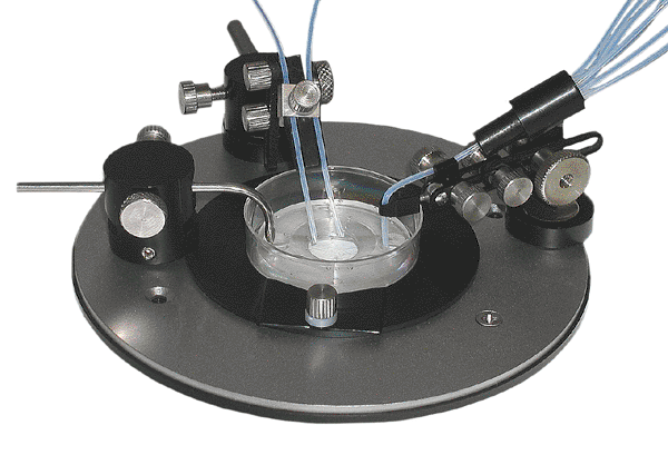
Low Profile Perfusion Set for Petri dishes, LPPCP1
Comes with PDI insert and 50
self-adhesive biocompatible gaskets,
which allow to use the assembly
with regular 35 mm Petri dishes.
Cells cultured in 35 mm Petri dishes are a popular research tool used in
numerous applications, including patch clamping and intracellular ion probe
imaging. However, true perfusion (continuous inflow and outflow) of solutions
can be difficult to configure. Drug delivery without an outflow requires a
spritzer-type microinjector, but ultimately results in contamination of the
entire dish after only a few applications. This forces scientists to plate cells
on cover slips for placement into specially designed perfusion chambers.
However, such transfer is a time consuming process which introduces the
potential for contamination plus additional expense.
The PDI chamber was
designed by scientists after several years of patch clamp research and calcium imaging combined with
external perfusion of single cells cultured in Petri dishes. The chamber has
separate openings for solution inflow and outflow that dump the fluctuations of
the liquid level in the working compartment and prevents bubbles
from entering the chamber. The laminar profile facilitates perfusion and provides faster solution exchange.
An adjustable metal suction tube (included) controls the level of liquid in the dish. The chamber was designed for use with
perfusion systems
and included magnetic holder with miniature ball-joint can accommodate
perfusion manifolds
(optional). Can be
used with glass bottom dishes
for imaging. In fact, some glass bottom dishes are made from standard Petri
dishes like Corning 35 mm, for example (Mattek dishes.) Can be used with temperature controlled systems.
The suction tubing requires connection to an outflow source CFPS-1U.
See published sample recordings,
and Sample publications.
Includes three magnetic tubing/electrode holders, and stainless
suction tubing. A magnetic microscope adapter
is required. Can be used with both magnetic and
non-magnetic microscope adapters.
If used with non-magnetic adapters, a IMA-MH set of miniature holders
is required.
Specifications: |
Outside diameter: | 35mm |
Height: | 3mm |
Working volume: | 11mm diameter, approx. 100 microl |
Click on catalog numbers below to purchase online.

Required accessories: microscope adapter,
miniature tubing holders.
Optional accessories: perfusion system,
temperature control,
flow control.
Download PDF manual.
Download PDF catalog.
Download PDF brochure.
|
|
Bioscience Tools
ph: 877-853-9755
fax: 866-533-7490
email: info@biosciencetools.com
|
PRICES AND OPTIONS
Petri
Dish Inserts |
LPPCP1 |
$475 |
Low Profile Perfusion System for Petri dishes.
|
MA |
$295 |
Magnetic microscope adapter, specify microscope model when ordering.
|
PDI |
$195 |
Replacement Low Profile Chamber-Insert for Petri dish |
IMA |
$195 |
Universal microscope adapters, specify microscope model when ordering.
|
IMA-MH |
$295 |
Miniature holders set for univesal adapters IMA, x3.
|
See published sample recordings,
and sample publications:
17
Enhancing Evanescent Wave Coupling of Near-Surface Waveguides with Plasmonic Nanoparticles.
Sensors 2023, 23, 3945;
16
Sites of Circadian Clock Neuron Plasticity Mediate Sensory Integration and Entrainment.
2021;
15
Evaluation of Optogenetic Electrophysiology Tools in Human Stem Cell-Derived Cardiomyocytes.
Frontiers in Physiology 8 October 2017;
14
A neural network underlying circadian entrainment and photoperiodic adjustment of sleep and activity in Drosophila.
Journal of Neuroscience, 36(35), 9084-9096. 2016;
13
Optogenetic Control of Gene Expression in Drosophila.
PLOS one, September 18, 2015;
12.
Analysis of functional neuronal connectivity in the Drosophila brain
J Neurophysiol. 2012 Jul 15; 108(2): 684–696;
11.
Differentially timed extracellular signals synchronize pacemaker neuron clocks.
PLoS Biol. 2014 September; 12(9);
10.
Functional Conservation of Clock Output Signaling between Flies and Intertidal Crabs.
Journal of Biological Rhythms, November 30, 2011;
10,
9,
8,
7,
6,
1,
2,
3,
4,
5.
|
|